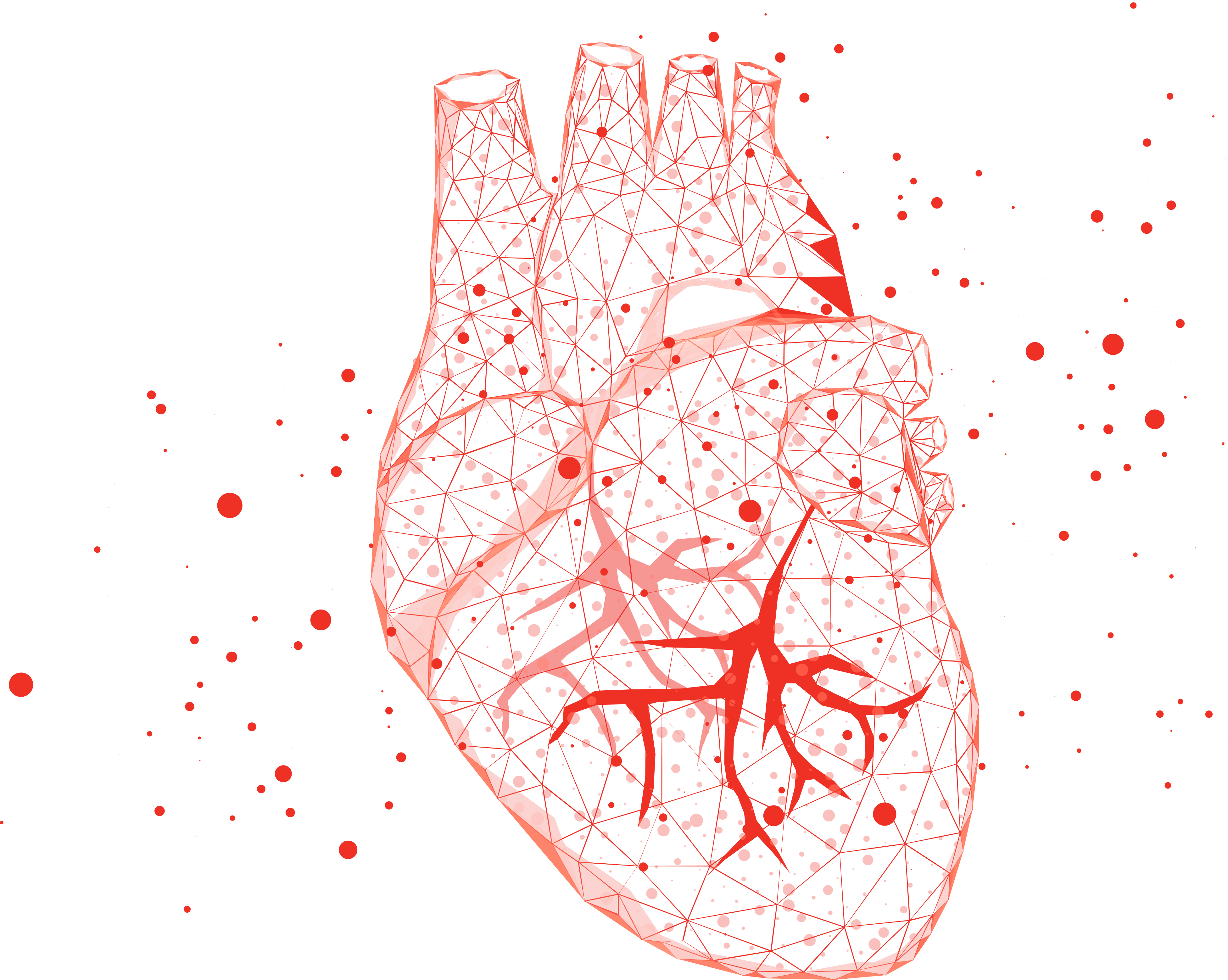
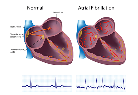
Atrial Fibrillation
Catheter Ablation
Electrical Cardioversion
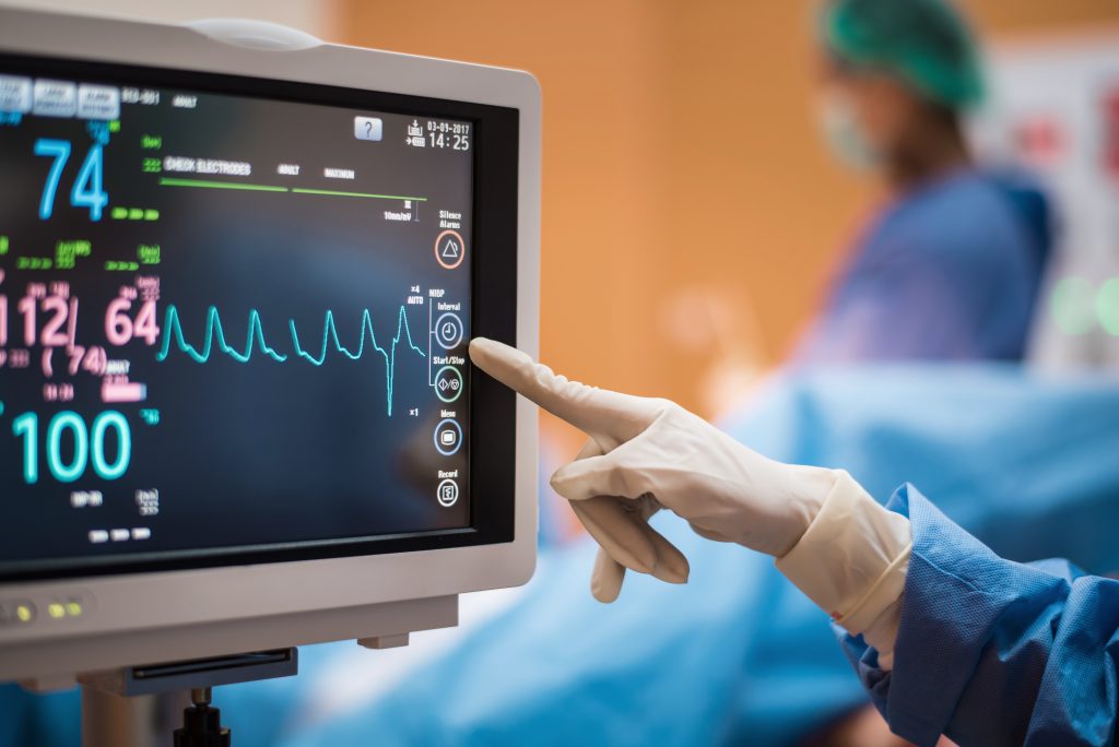
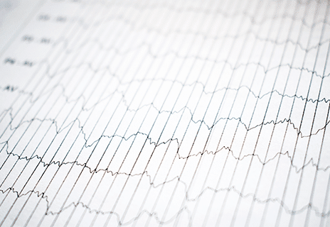
Electrocardiogram
Electrophysiology Study
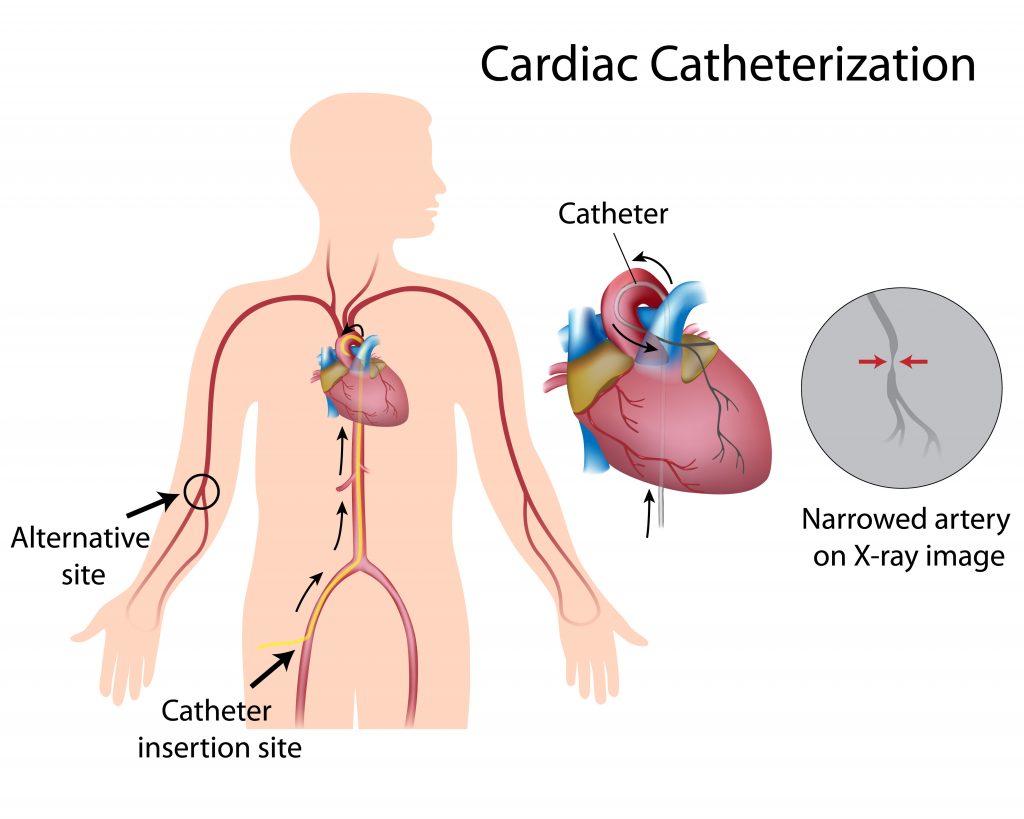
Expert Insights On Cardiac Catheterization
Heart Structure Tests

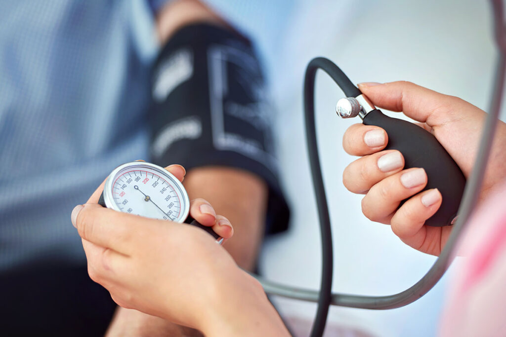
Hypertension
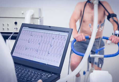
Stress Tests
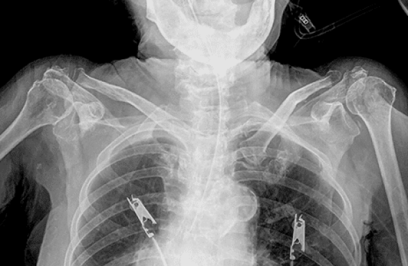
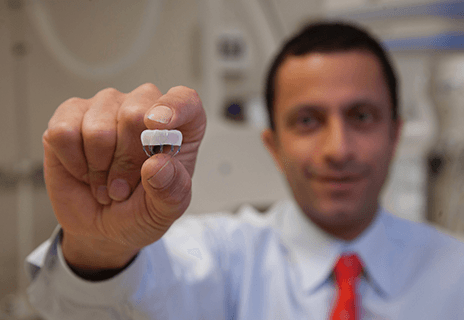
Watchman Implant Procedure
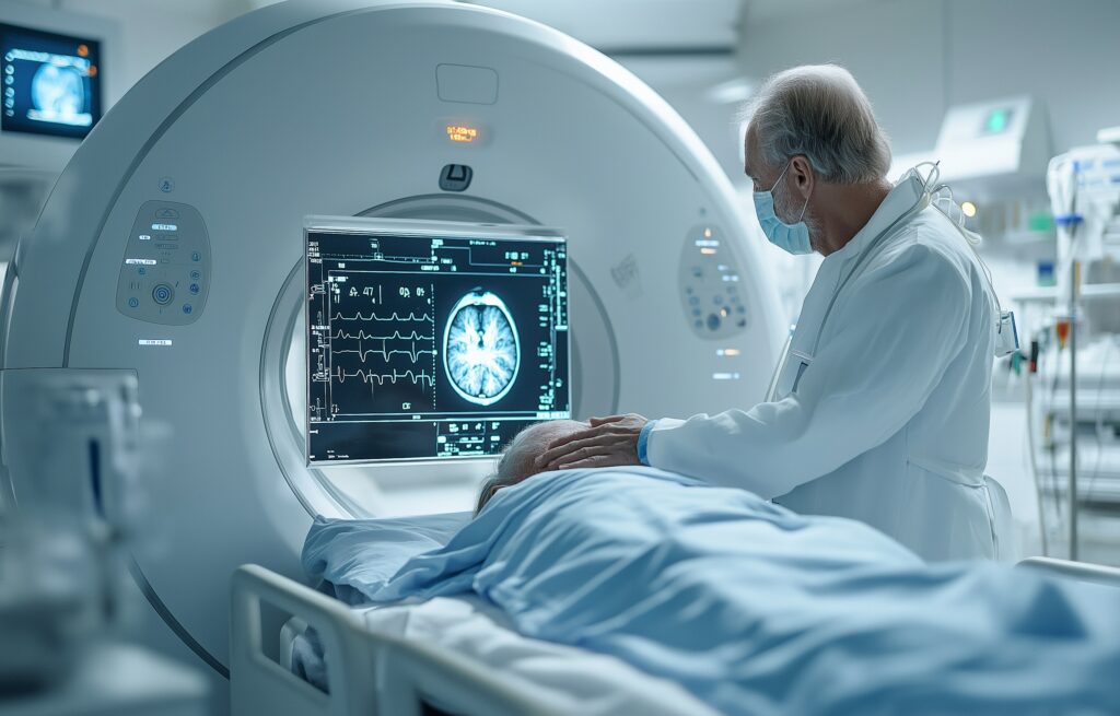
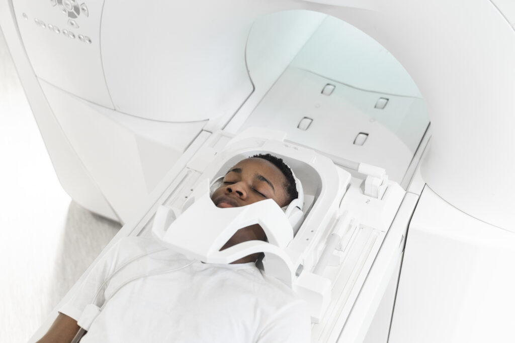
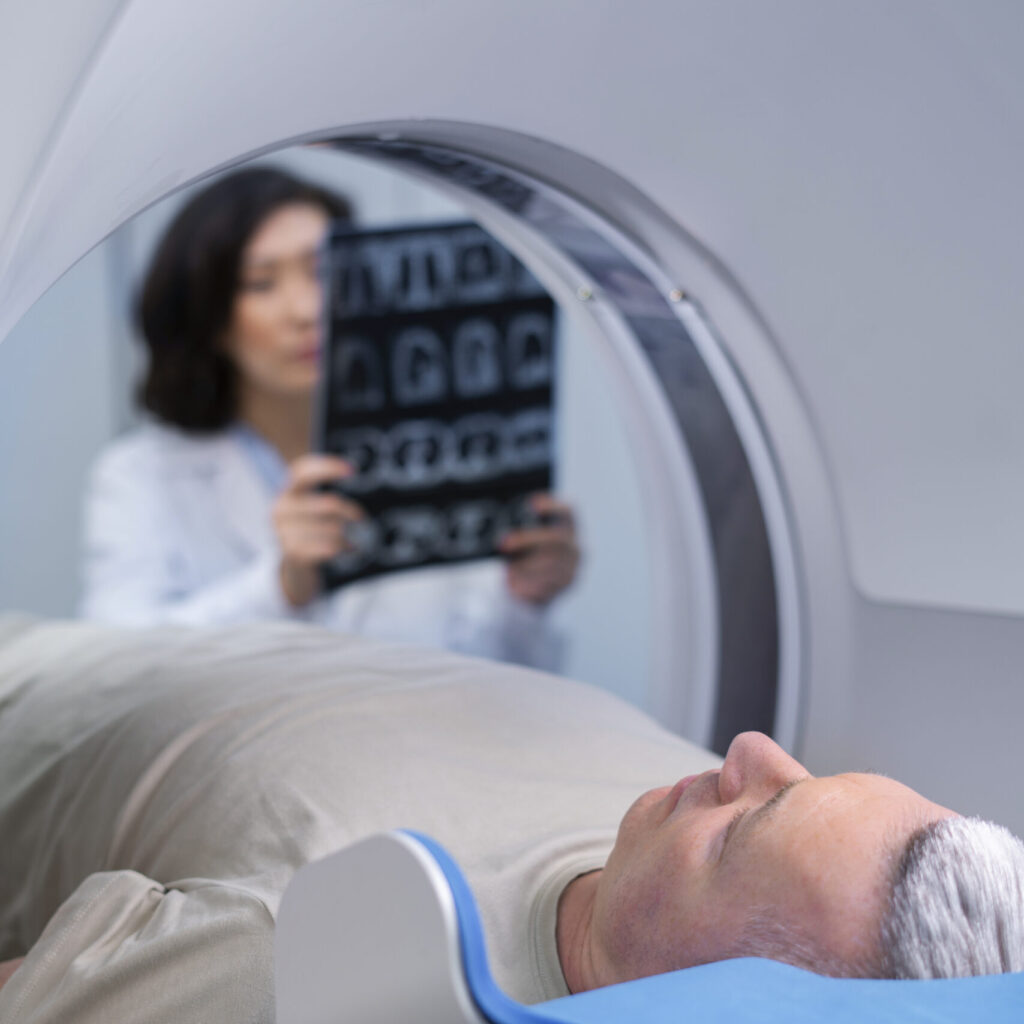
Cardiac CAT Scan
Cardio – Oncology
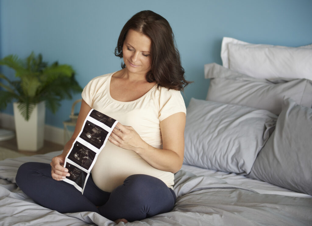
Cardio – Obstetrics
Cardiac MRI