|

  • |

  • If you received a bill or letter from Cardiovascular Anesthesiologist PC or Precision Anesthesia Billing, LLC please note that Cardiovascular Group and CVGcares.com is not affiliated with either of these organizations

    Cardiovascular Group and Northside Hospital are pleased to announce the signing of a Practice Services Agreement, signifying a major leap forward in cardiovascular patient care and clinical leadership in the Atlanta region.

    If your date of service is after June 1st 2021 use the button below to pay your bill online.


    Electrocardiogram

    Check For Signs Of Heart Disease With An Electrocardiogram

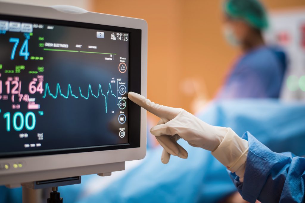
    An electrocardiogram, called an EKG or ECG, is a quick and painless test that records your heart’s electrical activity to check for signs of heart disease. A technician attaches multiple electrodes to your skin in various body places, tracking electrical impulses.

    What Can an EKG Check For?

    With an EKG, your doctor can check for multiple potential heart problems. These include irregular heart rhythms, poor blood flow, thickened heart muscle, electrolyte abnormalities, or even heart attacks. Any of these heart problems are risk factors for heart disease.

    Heart disease is a catch-all term that includes many types of heart problems, such as arrhythmias, pulmonary hypertension, and, most commonly, coronary artery disease (CAD). With an EKG, you can catch these early before they develop into more serious issues like heart failure.

    How Does an EKG Work?

    A technician attaches multiple electrodes to the skin on your chest, arms, and legs. During a resting EKG, you’ll lie flat while a computer records your electrical impulses moving through your heart, creating a picture of the activity. A stress test can also be conducted, where the same test is administered to check your heart rate while you exercise. Besides the standard EKG tests, other kinds of tests can also be used to test your heart.

    A Holter monitor is a portable EKG that monitors the electrical activity of your heart 24/7 over 1 to 2 days. This test may be suggested if your doctor suspects you have an irregular heart rhythm, heart palpitations, or low blood flow. You can go about your daily routine except for showering during this test. You’ll record your activities and any symptoms you notice during this time.

    An event monitor is a less intensive test for occasional symptoms. This device has a button you can press whenever you notice symptoms, recording and storing your heart’s electrical activity for a few minutes. Because of the low frequency of this test, you may need to wear the device for weeks or even months.

    A loop recorder is a device implanted into your body under the skin of your chest. It has the same functions as an EKG but allows continuous remote monitoring of your heart’s electrical activity. This device searches for irregularities that cause more severe symptoms, such as fainting and heart palpitations.

    What Symptoms Should You Get Checked with an EKG?

    If you have any of the following symptoms, it may be time to get an EKG test:

    • Chest pain
    • Dizziness or lightheadedness
    • Heart palpitations
    • Shortness of breath
    • Rapid pulse
    • Weakness or fatigue
    • Inability to exercise

    If your family has a history of heart disease, your doctor may suggest an electrocardiogram even if you aren’t displaying any symptoms.

    Risks Of An EKG

    There are little to no risks involved with EKG testing. Some people with skin sensitivity can experience a rash where the electrodes are placed; however, this usually goes away independently. Patients who undergo the stress test may be at risk of a heart attack, although this is related to the exercise and not the EKG itself.

    An EKG only monitors the electrical activity of your heart; it does not emit any electricity. This means the device is completely safe, even during pregnancy.

    How To Prepare for an EKG

    While getting ready for your EKG, there are a few things to consider:

    • Remove any jewelry
    • Shave hair in areas where the electrodes will be placed
    • Avoid drinking cold water directly before the test (this can cause changes in electrical patterns)
    • Do not participate in any exercise or other activity that will increase heart rate before the test
    • Keep the room at a moderate temperature to avoid shivering (movement will affect the accuracy of the results)

    Results Of An EKG

    Your EKG results can sometimes be available the same day as your test or by the next appointment. The results of an EKG will give your doctor more details about the following issues:

    • Heart rate: Heart rate can typically be measured by checking your pulse. An EKG is helpful if your pulse is irregular, too fast, too slow, or at odd moments. In these instances, your pulse will be difficult to determine regularly. An EKG can help determine an abnormally fast heart rate (tachycardia) or an unusually slow heart rate (bradycardia).
    • Heart rhythm: An EKG can determine if you have an irregular heartbeat (arrhythmias). Arrhythmias can occur if parts of the heart’s electrical system aren’t working properly.
    • Heart attack: An EKG can show evidence of previous heart attacks or a current one. It helps to give detailed results on which part of the heart has been damaged and the extent of the damage.
    • Blood and oxygen supply: An EKG done while experiencing symptoms will help your doctor determine if chest pain is caused by reduced blood flow to the heart.
    • Heart structure: An EKG can provide doctors with more information about whether you have an enlarged heart, heart defects, or other heart issues.

    How CVG Can Help

    CVG offers electrocardiograms to assess your risks of heart disease. We provide various EKGs so that we can determine how your heart is functioning and what steps you will need to take to prevent further problems. If these tests determine a problem, we offer treatment solutions to help against heart disease.

    Learn more about our services here, or schedule an appointment to talk to our doctors.

    Back to services
    Electrocardiogram

    Frequently Asked Questions

    If there is an acute emergency, the best thing to do is immediately contact 911. If you think you may be having a heart attack, also immediately call 911. When the paramedics arrive, or you are seen in the emergency room, you should notify them who your cardiologist is so they can directly notify our physicians.

    Yes. We have physicians available 24 hours a day for acute care needs. A physician from our group is also always available to perform an emergency procedure for an acute heart attack. Please let the paramedics or emergency room staff know who your cardiologist is so they can notify the physician on call.

    Yes. Physicians are available every weekend and holiday to care for all your inpatient needs. This may not be your primary cardiologist; however, he has informed the on-call physician about your current in-hospital stay, and outpatient records are always available if needed.

    Yes. After every visit, a letter is dictated and sent to your primary care physician. We are also in frequent telephone contact with them. Our physicians also work closely with your primary care physician in the hospital setting if you are hospitalized. Your cardiologist can recommend one if you do not have a primary care physician. If you switch primary physicians, please notify our staff so we can arrange to have further evaluations sent correctly.

    We offer a complete blood lab in our office. If we cannot accommodate a specific lab test for you, our front office staff will happily arrange it.

    If you need a refill of your heart medicines, please contact our medical assistants or log on to your account through the Patient Portal (here). They will then notify your physician, who can authorize refills for you. When you see your physician in the office, bringing your medications with you is always best. Our medical assistants will update your chart and notify the doctor if drugs must be reordered.

    No. While we work closely with many primary care physicians in the community, you do not need a referral to have an evaluation with our physicians.

    We strive to be as accessible as possible to our patients. In addition to participating in most insurance plans in our area, we offer flexible payment plans and accept MasterCard, Visa, American Express, and Discover.

    If you have any questions about our participation in a particular plan or network, call us at 770-962-0399.

    If this is the case, please get in touch with our business department. They can often assist you with a payment program tailored to your needs.

    Unfortunately, it can be confusing for a patient when experiencing chest pain, especially if it's on the left side. The concern is whether it's related to the heart. While we know the typical description of heart chest pain, it doesn't always conform to textbook explanations. It can feel like indigestion, with burning sensations, not necessarily on the left side but also behind the breastbone or on the right side. Heart pain due to lack of blood supply doesn't usually last for a few seconds; it often occurs during physical exertion.

    There are different characteristics and precipitating factors that we, as physicians, ask the patients when trying to determine whether the chest pain is truly due to a lack of blood to the heart. There are multiple other causes of chest pain. Concerning the heart, one can also experience chest pain secondary to inflammation of the outer sac of the heart, called pericarditis. This pain is typically worse when one lies down and better when sitting up. The most common cause is a viral syndrome. Therefore, it is usually preceded by a fever, cold, or flu-like symptoms.

    Chest discomfort post-meal or lying down is often from indigestion or heartburn—a burning sensation behind the breastbone that may extend to the jaw. Gallbladder pain, especially after fatty meals, is typically on the right side and may spread to the shoulder blade, often with nausea. Pain below the breastbone or left side of the upper abdomen that eases with eating is likely stomach inflammation or an ulcer. Chest pain worsening with deep breaths or coughing may stem from a lung issue like pleurisy, often caused by a viral infection. Superficial chest wall pain from muscle, bone, or nerve injury can occur. In females, chest pain from the breasts can mimic heart-related pain.

    An EKG is a snapshot of the electric patterns in your heart that make it beat. It is a routine test in any cardiac evaluation. You can expect one during an initial assessment and any time symptoms change. It is also used to monitor the effects of certain medications. A medical assistant or nurse in the office administers the test. It allows us to look for irregularities in the electrical rhythm of your heart and gives us clues to structural abnormalities of the heart.

    Heart failure occurs when the heart cannot pump efficiently to circulate oxygen-rich blood to different body organs. Simply put, the heart cannot keep up with its workload.

    - Shortness of breath

    - Easy fatigue

    - Coughing, particularly when lying down

    - Swelling in the ankles

    - Weight gain

    - Loss of appetite

    - Rapid heartbeat

    - Difficulty tolerating exercise

    Common causes of heart failure include:

    - Heart muscle weakness (Cardiomyopathy)

    - Stiffness of the heart

    - Valvular abnormalities such as leaky or narrowed valves

    - Issues with the heart's electrical system (Arrhythmias)

    - Arterial blockages and heart attacks

    - Hypertension

    - Congenital heart defects

    - Infections

    - Obesity

    - Nutritional deficiencies

    Treatment for heart failure varies based on the cause and symptoms. Lifestyle changes can reduce signs, sometimes even reversing heart failure causes. Medications such as ACE inhibitors, beta-blockers, angiotensin receptor blockers, diuretics, and antiarrhythmics are common. Implanting devices like pacemakers, defibrillators, or an artificial heart (LVAD) may be necessary for severe cases. In refractory instances, a heart transplant could be considered for certain patients.

    Preventing heart failure hospitalizations involves key steps:

    - Adhering to prescribed medications consistently.

    - Monitoring weight regularly for any sudden gain exceeding 2 pounds daily.

    - Notifying your doctor of increased shortness of breath, lower limb swelling, or notable weight gain.

    - Adopting a low-salt diet.

    - Manage blood pressure and blood sugar levels and address underlying issues like sleep apnea.

    To prevent heart failure, consider these steps: Embrace dietary adjustments such as opting for a low-salt diet. Avoid smoking and alcohol abuse, and prioritize physical activity—address underlying conditions like high blood pressure, diabetes, sleep apnea, and arrhythmias. Manage stress effectively, as it can elevate blood pressure and heighten the risk of heart attacks.

    The outlook for individuals with heart failure can vary significantly based on factors such as the cause, risk factor management, lifestyle adjustments, and adherence to prescribed medications. If left untreated or if underlying issues are unaddressed, heart failure can deteriorate over time, potentially leading to a fatal outcome. It is crucial to consult your healthcare provider to explore tailored treatment options.

    Various heart diseases can increase the risk of sudden cardiac arrest. A cardiac arrest typically occurs due to an electrical storm in the ventricles, the heart's main pumping chambers. When this occurs, the heart essentially stops beating, and if left uninterrupted, it results in almost immediate death. An implantable defibrillator (or ICD) can protect you.

    Implantation closely resembles pacemaker insertion. The ICD is usually placed under the skin via a small incision beneath the collarbone. A lead is then guided through a vein to the heart, making contact with its lining. This lead enables the defibrillator to monitor the heartbeat. Upon detecting any cardiac electrical irregularities, the defibrillator can charge and deliver a shock to reset the heart, akin to having a paramedic ready. The procedure is performed under sedation and local anesthesia. You may be discharged on the same day or require an overnight stay, depending on the circumstances.

    After the implantation, you may experience minimal pain and slight swelling at the site. Pain medication will be provided if needed. Expect to wear a sling for a few days, avoid lifting more than 10 pounds, and keep your elbow below shoulder level for 3 weeks. Steer clear of strenuous activities like a full golf swing for several months. Follow-up appointments are typically scheduled in the next couple of weeks, with regular check-ins at our device clinic once or twice a year. Home monitoring can complement these office visits.

    Pacemakers are used to prevent a slow heartbeat. A slow heartbeat is caused either by the natural pacemaker of your heart, the sinus node, firing too slowly or because those electrical impulses don't make it reliably from the upper chambers, the atria, the lower chambers, and the ventricles.

    A pacemaker is typically implanted beneath the skin through a small incision below the collarbone. Pacemaker leads are then threaded through a vein to the heart, where they touch the heart's lining. These wires allow the pacemaker to sense the heartbeat and stimulate the heart to beat if it is too slow. The procedure is done with sedation and local anesthetic. Depending on the situation, you may be able to go home the same day or may need to stay overnight.

    After implantation, the site usually has minimal pain and mild swelling. Pain medication will be provided if needed. A sling is worn for a few days. For three weeks, avoid lifting over 10 lbs. and keep the elbow below shoulder level. Refrain from vigorous activity like a full golf swing for a few months. Follow-up appointments are typically scheduled within a couple of weeks. Pacemakers are usually monitored in our device clinic once or twice a year, with the option of home monitoring for additional checks.

    The main pumping chamber of the heart is the left ventricle. However, the effectiveness of that pump is compromised if the sides of the left ventricle don't squeeze simultaneously. This lack of synchronization affects the heart's output and can lead to progressive weakening and enlargement of the heart. When that lack of synchronization is caused by a delay in the electrical signal directing the heartbeat, it can be corrected by a special pacemaker known as a biventricular pacemaker. This is also referred to as cardiac resynchronization therapy. This pacing technique can be applied to both pacemakers and implantable defibrillators.

    During either pacemaker or defibrillator implantation, an extra “third wire,” also known as a left ventricular lead, is placed. This is threaded to the heart the same way as other pacemaker or defibrillator leads, except it is then further threaded from the right atrium into another vein that leads to the surface of the left ventricle. Getting that third wire in place is trickier but achievable in about 95% of cases. Otherwise, the rest of the procedures are the same as a standard pacemaker or defibrillator implantation. Please see the descriptions of those procedures.

    Please see the details for the defibrillator or pacemaker implantation. However, unique to biventricular pacing, the left ventricular lead passes very close to a nerve that runs along the surface of the heart from the spine down to the diaphragm. This is called the phrenic nerve. If the left ventricular leave stimulates it, it can cause a twitch of the left side of the diaphragm with every heartbeat. This is not dangerous but can be quite aggravating. It can usually be corrected by reprogramming the pacemaker or defibrillator in the office. We frequently watch people overnight because of the potential for phrenic nerve stimulation.

    Heart disease is the primary cause of death in the United States, accounting for 1 in 4 female deaths. The number of women and men succumbing to heart disease each year is equal. Surprisingly, two-thirds of women who passed away suddenly from heart disease had no prior symptoms. Approximately 6% of white women, 8% of African-American women, and 6% of Mexican American women are affected by coronary artery disease.

    More women present with atypical symptoms than men, and some women have no symptoms of heart disease. Common Heart attack symptoms in women can be pressure in the chest, shortness of breath, jaw pain, throat pain, and pain in the upper abdomen or back. Pain can be sharp, dull, burning, or indigestion-like. These episodes can occur during rest or physical activity or can be triggered by mental stress.

    Hypertension. High cholesterol. Smoking. Diabetes. Excessive alcohol use. Physical inactivity. Overweight and obesity. Poor diet.

    Women usually develop heart disease a decade later than men. Microvascular disease, impacting the heart's small arteries, is more common in women. Mitral valve prolapse is also more frequent in women. Additionally, women are more prone to conditions like broken heart syndrome, where extreme stress can cause heart muscle failure and palpitations. By modifying risk factors, changing lifestyles, using medical treatments, and considering surgeries, women can decrease heart disease mortality risk and improve symptoms like men. Early prevention, accurate diagnosis, and timely treatment are crucial for better survival rates.

    Have A Question?
    • This field is for validation purposes and should be left unchanged.
    678-582-8586
    Request An Appointment