
Cardiac MRI - Advanced Heart Imaging at CVG
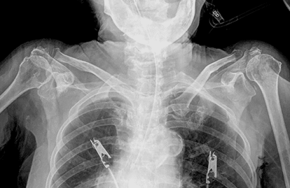
Quick, painless chest X-ray services ordered and interpreted by CVG’s board-certified cardiologists, available across Gwinnett County and the Atlanta metro.
A chest X-ray is one of the most common and valuable diagnostic tools in cardiovascular medicine. It uses a small, controlled amount of radiation to produce clear images of your heart, lungs, aorta, and surrounding blood vessels, giving your CVG cardiologist a fast, non-invasive look at your cardiovascular health without the need for more complex testing.
At CVG, chest X-rays are ordered and interpreted by our board-certified cardiologists as part of a comprehensive cardiac evaluation. Whether your doctor has noticed an abnormality during a routine exam, you’re experiencing symptoms such as shortness of breath or chest pain, or you need baseline imaging to monitor a known heart condition, a chest X-ray at CVG provides clinically meaningful information quickly and comfortably.

What is a Chest X-ray?
Some patients need multiple chest X-rays so physicians can obtain a baseline and track conditions over time. X-rays are a convenient, efficient imaging technique that provides life-saving information to doctors. Thankfully, chest X-rays expose patients to only a minimal amount of radiation, which benefits those who need one. Overall, a chest X-ray is quick, easy, and painless.

What Does A Chest X-Ray Reveal?
Heart size and shape
An enlarged heart (cardiomegaly) may indicate heart failure, cardiomyopathy, or fluid around the heart
Fluid in or around the lungs
A sign of congestive heart failure or pericardial effusion
Aortic abnormalities
Changes in the size or contour of the aorta that may suggest an aneurysm or dissection
Calcium deposits
Calcification in the coronary arteries, heart valves, or aorta is visible on X-ray
Pulmonary vascular changes
Pacemaker and device positioning
Confirming correct placement of implanted cardiac devices including pacemakers, defibrillators, and catheters
Lung conditions with cardiac implications
What to Expect During Your Chest X-Ray at CVG
A chest X-ray is one of the quickest and most straightforward tests in cardiovascular medicine. Here is what to expect:
Before the X-Ray: No special preparation is required for a standard chest X-ray. You will be asked to remove jewelry, piercings, and any clothing with metal fastenings from the chest area. If you are pregnant or think you may be pregnant, inform your CVG care team before the X-ray. While radiation exposure is very low, your physician may recommend an alternative imaging approach.
During the X-Ray: The X-ray itself takes less than a minute. You will stand in front of the X-ray machine and be asked to take a deep breath and hold it briefly, typically for just a few seconds, while the image is captured. Most chest X-rays require two views: one from the front and one from the side. The process is entirely painless and involves no injections or inserted equipment.
After the X-Ray: There is no recovery time. You can resume normal activities immediately. Your CVG cardiologist will review the images and discuss the findings with you, explaining what is normal, what may require further evaluation, and what next steps are recommended for your cardiovascular care.

Normal vs. Abnormal Chest X-Ray Results
Normal Chest X-Ray Results
A normal chest X-ray shows a heart that is the expected size and shape for your body, clear lung fields with no signs of fluid or infection, and well-defined blood vessels and aorta. A normal result provides a valuable baseline that your CVG cardiologist can reference for comparison at future evaluations.
Abnormal Chest X-Ray Results
An abnormal chest X-ray may reveal findings such as an enlarged heart, fluid around or within the lungs, aortic enlargement, calcium deposits, or changes in lung structure. An abnormal result does not always mean a serious problem, but it does mean your cardiologist needs to investigate further. At CVG, if your chest X-ray reveals an abnormality, your physician will review the findings with you in detail and recommend the appropriate next steps, which may include additional imaging such as an echocardiogram or cardiac MRI, or further blood work.
Frequently Asked Questions About Chest X-Rays
A normal chest X-ray shows a heart of expected size and clear lung fields with no fluid or structural abnormalities. An abnormal chest X-ray may show an enlarged heart, fluid in the lungs, aortic changes, or other findings that warrant further evaluation. Your CVG cardiologist will explain any abnormal findings and recommend appropriate next steps.
The X-ray itself takes less than one minute. Including preparation and positioning, most patients are in and out within 15 to 20 minutes. There is no recovery time required.
Yes. The radiation exposure from a chest X-ray is very low, equivalent to a small fraction of your natural daily background radiation exposure. Chest X-rays are considered safe for most adults. If you are pregnant or may be pregnant, inform your CVG physician before the test.
A chest X-ray at CVG is ordered by your cardiologist as part of your evaluation. Contact your nearest CVG location to schedule a consultation, and your physician will determine whether a chest X-ray is the appropriate next step for your care.
Why Choose CVG
CVG’s cardiologists don’t just order chest X-rays; they interpret your results as part of your complete cardiovascular picture. If your X-ray reveals an enlarged heart, fluid in the lungs, or another abnormality, your CVG physician will explain what it means, recommend any additional testing needed, and work with you on a clear next step for your heart health.
With 11 locations across Gwinnett County and the Atlanta metro and a track record of caring for Georgia patients since 1982, CVG makes expert cardiac imaging accessible without the wait of a hospital system referral.
Lawrenceville (Main Office)
Buford
Loganville
2200 Medical Center Blvd, Suite 400
Lawrenceville, GA 30046
770.962.0399 | 770.290.8084
2800 Buford Drive, Suite 320
Buford, GA 30519
770.962.0399 | 770.290.8084
98 Tara Commons
Loganville, GA 30052
770.962.0399 | 770.290.8084